Hi Mli,
Thanks for the photos. Would you be able to also provide photos showing both lateral (side) views?
Follow the good advice given to continue fridging your axolotl. The condition definitely requires a vet for full diagnostic work up.
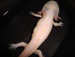
Impaction is a possibility and radiographs will be able to pick up some forms of mineralised obstruction. However there are other causes of lesions and herniation similar to that clinical presentation as well as other obstructions that may not be picked up radiographically. Some tumours or neoplasia especially if affecting the peritoneal (abdominal) cavity (Inside out) may not show up on radiographs but can show the protrusion externally on the skin.
A bacterial infection can also cause lesions to appear like that, although most aquatic species get gram negative type infections more commonly, abscesses and granulomas can still develop. Some types of fungal infection, embedded foreign body (such as from a previous trauma) as well as bacteria like mycobacteria can all cause granulomas to form. They can take on that irregular, poorly circumscribed nodular lesion as seen the photos.
Your vet might perform a fine needle aspirate to obtain some cellular material from the lesions to look under the microscope. Staining and cytology will help distinguish the underlying cause, such as neoplasia (abnormal cells), abscess (inflammatroy cells) or chronic granuloma (fiibrous tissue).
Cheers